Fig. 5
Download original image
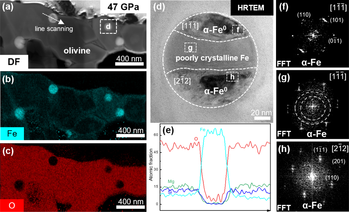
TEM images of iron particles. (a) DF image of olivine shocked at 47 GPa, showing nanoscale particles concentrated at the corners of olivine grains. The dashed arrow marks the location of the elemental line scan, while the dashed box marks the area corresponding to image (d). (b and c) Fe and O elemental mapping of olivine shocked at 47 GPa, revealing that the nanoscale particles are iron-rich and oxygen-free. (d) HRTEM image of an iron particle, with a diameter reaching up to 100 nm. The particle exhibits distinct contrast variations, divided into upper, middle, and lower regions, which are identified as α-Fe through FFT calibration. (e) Elemental line scan across the iron particle, demonstrating a sharp increase in Fe abundance, while the O, Mg, and Si abundances drop to zero within the particle. (f–h) FFT patterns of the upper, middle, and lower regions of the iron particle. The upper region has a crystal orientation of ![]() , the middle region exhibits an amorphous ring alongside a crystal orientation of
, the middle region exhibits an amorphous ring alongside a crystal orientation of ![]() , and the lower region displays a crystal orientation of
, and the lower region displays a crystal orientation of ![]() .
.
Current usage metrics show cumulative count of Article Views (full-text article views including HTML views, PDF and ePub downloads, according to the available data) and Abstracts Views on Vision4Press platform.
Data correspond to usage on the plateform after 2015. The current usage metrics is available 48-96 hours after online publication and is updated daily on week days.
Initial download of the metrics may take a while.