Fig. 2.
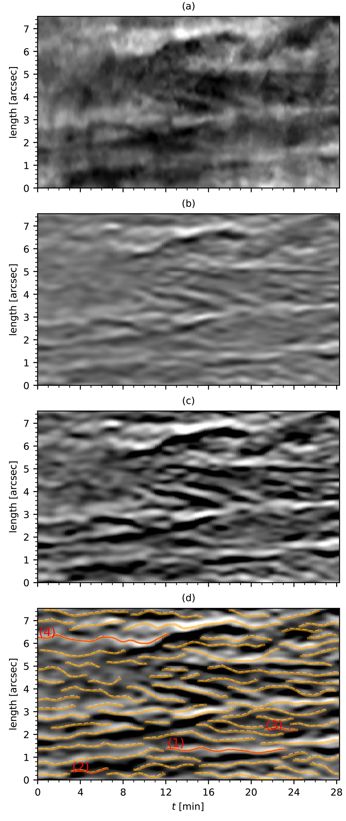
Download original image
Time evolution of the cut across the bright fibrils shown with a dashed white line (Fig. 1f). Panel (a) displays the intensity at the nominal line centre of Ca II K. Panel (b) shows the enhanced intensity of panel (a) by applying the enhancing procedure described in Sect. 3.1. Panel (c) is the gamma-corrected image of panel (b) that fully brings out individual fibril oscillations. The POS oscillation trajectories are marked on the high contrast space-time image in panel (d), where the preliminary paths are plotted with dashed lines and the smoothed oscillations are plotted with solid curves. The oscillation properties of the curves marked with numbers are shown in Fig. 8. The zero point of the cut length (y-axis) is marked with a white dot in Fig. 1f.
Current usage metrics show cumulative count of Article Views (full-text article views including HTML views, PDF and ePub downloads, according to the available data) and Abstracts Views on Vision4Press platform.
Data correspond to usage on the plateform after 2015. The current usage metrics is available 48-96 hours after online publication and is updated daily on week days.
Initial download of the metrics may take a while.