Fig. 1.
Download original image
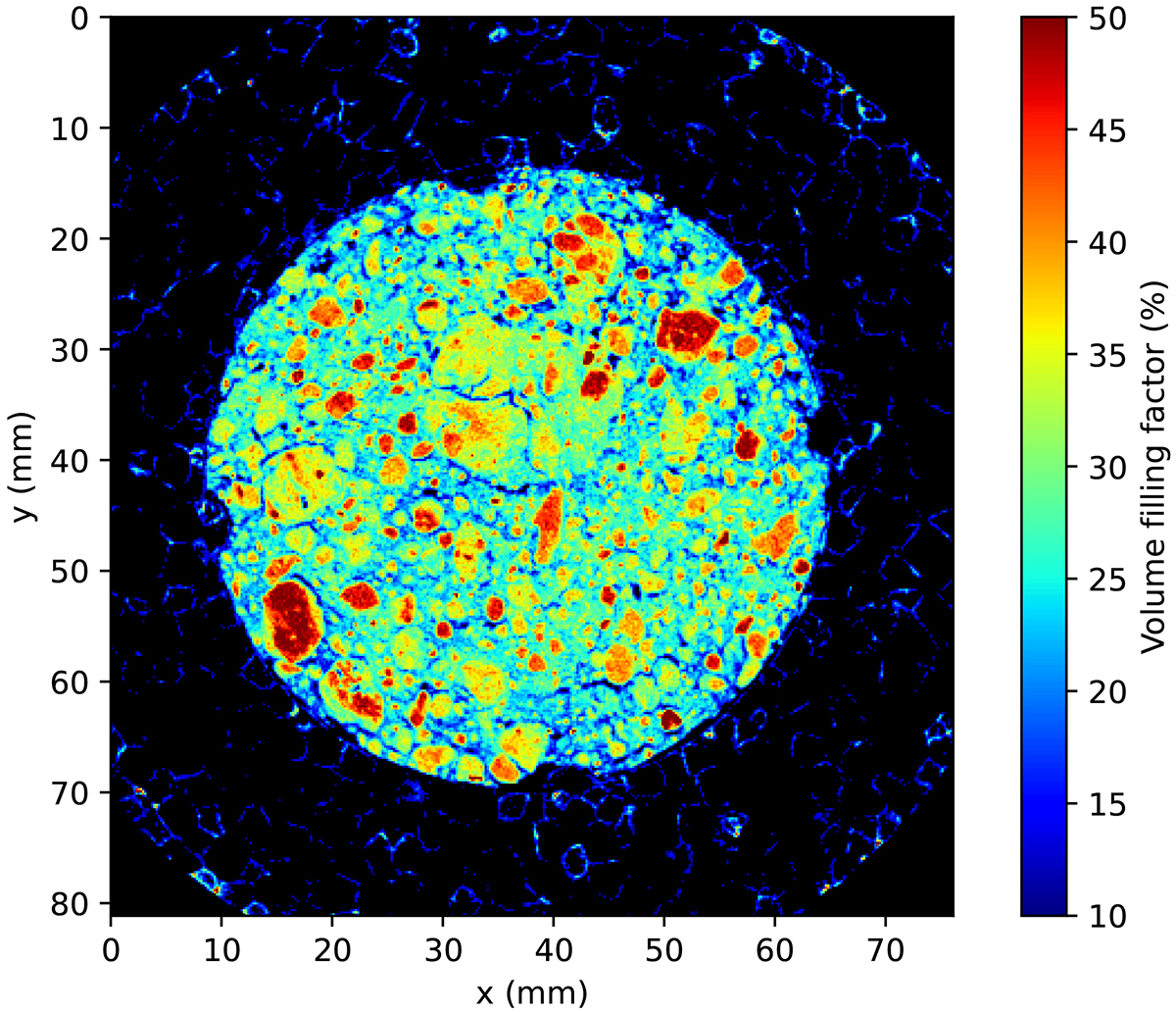
One of the reconstructed images of the CT scan of the spooned sample, showing its inner structure dominated by pebbles of various sizes. The voxel size of the CT scan is 67 μm. The selected slice was taken from the middle of the sample and shows the cylindrical granular water ice surrounded by the cylindrical styrofoam container used as a sample holder during the scans to keep the sample cold. This sample holder has four bulges extending into the sample, as visible in the image. The structure of the styrofoam is visible in the image; however, the colour bar on the right applies only to the water-ice sample. The corners of the image were filled with air during the CT scan and were used as zero-density analogues for the calibration of the scans. The other slices and the poured sample are similar in appearance.
Current usage metrics show cumulative count of Article Views (full-text article views including HTML views, PDF and ePub downloads, according to the available data) and Abstracts Views on Vision4Press platform.
Data correspond to usage on the plateform after 2015. The current usage metrics is available 48-96 hours after online publication and is updated daily on week days.
Initial download of the metrics may take a while.