Fig. 6
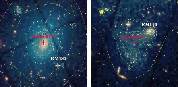
Two problematic matching cases, which were a posteriori recovered by visual inspection. The SDSS colour 7′ × 7′ image is overlaid on the raw X-ray photon image + contours (the X-ray image is not corrected for detector cosmetic); the optical and X-ray centres are marked by a white and a red cross, respectively. Left: wrong optical centre; the offset between the redMaPPer and X-ray centres is 2.2′. Right: complex nearby structure; the offset between the two centres is 1.7′.
Current usage metrics show cumulative count of Article Views (full-text article views including HTML views, PDF and ePub downloads, according to the available data) and Abstracts Views on Vision4Press platform.
Data correspond to usage on the plateform after 2015. The current usage metrics is available 48-96 hours after online publication and is updated daily on week days.
Initial download of the metrics may take a while.