Fig. 1
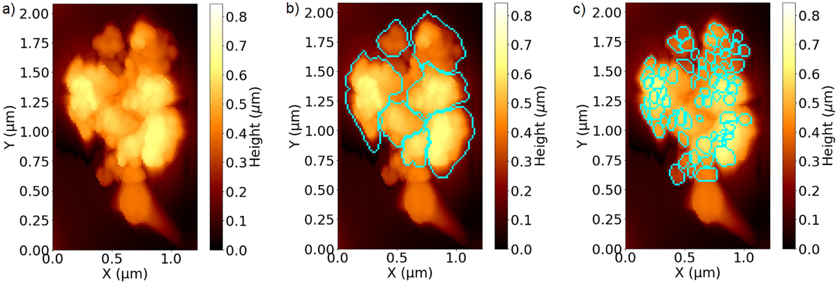
Particle G, a 1 μm particle scanned with the MIDAS reverse-imaging mode on 8 December 2015 with a resolution of 15 nm per pixel. The smooth, round shape at the bottom with a fading line to the bottom right corner is the tip with which the particle was picked up, above sits the particle with well-visible substructures. Panel a: particle itself. Panel b: larger subunits. Panel c: smallest identifiable features.
Current usage metrics show cumulative count of Article Views (full-text article views including HTML views, PDF and ePub downloads, according to the available data) and Abstracts Views on Vision4Press platform.
Data correspond to usage on the plateform after 2015. The current usage metrics is available 48-96 hours after online publication and is updated daily on week days.
Initial download of the metrics may take a while.