Fig. 2
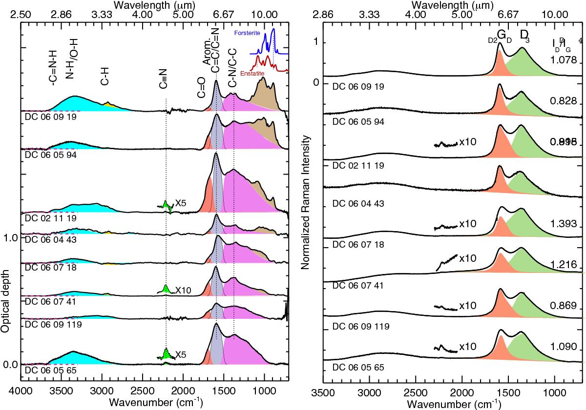
Left: UCAMM samples μFTIR optical depth spectra. The spectra have been vertically shifted for clarity (left horizontal dashed lines indicate the shift level). They are ordered from top to bottom by decreasing amount of silicate band absorption. The main band contributions are labelled above the upper spectrum. The spectra are deconvolved into several contributions shown with distinct colours, used in the analysis. A zoom on the nitrile region (~2200 cm-1) is provided when a band was measured. Right: UCAMM samples Raman spectra, normalized to the G-band maximum. The spectra are analysed using a classical Raman bands fitting procedure (Sadezky et al. 2005; Kouketsu et al. 2014) contributing to the D (green) and G (red) bands. The D/G band peak intensity ratio is given on the right, as defined in Busemann et al. (2007). A zoom on the nitrile region (~2200 cm-1) is provided when a band was observed. Spectra are vertically shifted for clarity.
Current usage metrics show cumulative count of Article Views (full-text article views including HTML views, PDF and ePub downloads, according to the available data) and Abstracts Views on Vision4Press platform.
Data correspond to usage on the plateform after 2015. The current usage metrics is available 48-96 hours after online publication and is updated daily on week days.
Initial download of the metrics may take a while.