Fig. 6
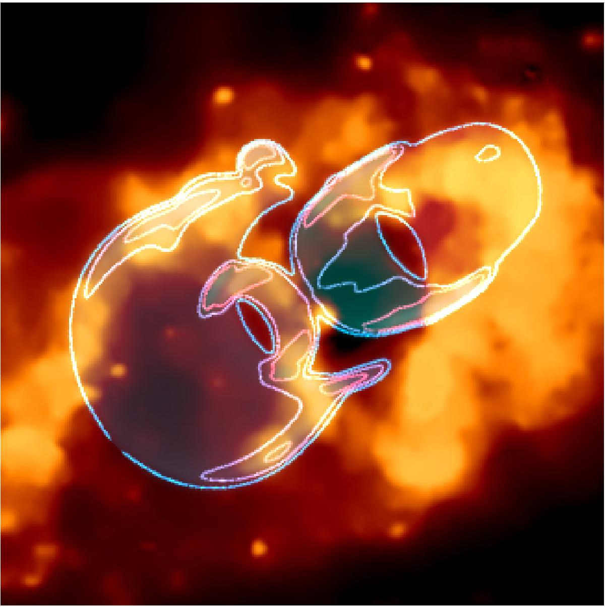
Composite images displaying our SHAPE model together with the 0.5–1.2 keV Chandra X-ray image. The soft X-ray emission is produced as material runs into the nearby gas and dust. X-ray emission from the Homunculus is detected only in hard X-ray bands. The large-scale optical tube structure fits right inside the X-ray bubble. The SHAPE model contours only qualitatively show the outlines of the emission. The blue and red colors are to distinguish the different shapes. There is no physical information associated with them. For the image scale, see Fig. 7.
Current usage metrics show cumulative count of Article Views (full-text article views including HTML views, PDF and ePub downloads, according to the available data) and Abstracts Views on Vision4Press platform.
Data correspond to usage on the plateform after 2015. The current usage metrics is available 48-96 hours after online publication and is updated daily on week days.
Initial download of the metrics may take a while.