Fig. 2
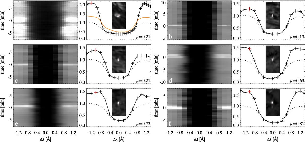
Sample λ−t diagrams and spectral profiles of Ellerman features in Hα at the observing angles specified at lower-right in the profile plots. Panel pair a) is from dataset 2, b) and c) from 1, d) from 8, e) from 7, f) from 6. Pair a) shows an active-region EB for comparison, the one at the bottom at (x,y) ≈ (29″,15″) in Hα-panel D of Fig. 1. The other pairs show QSEBs. Each λ−t image is greyscaled between 0.3 and 1.5 times the Hα far wing intensity. The Ellerman feature occurs at time 0. The profiles are normalized to the outer-wing intensity of the time-averaged profile (dashed) of an area close to the Ellerman feature. Note the scale difference for profile graph a). The orange profile added to a) is from the facular brightening visible at the top of the small inset image. The red pluses in the blue wing of each feature profile (solid) specify the spectral sampling of the small image insets (size 2.3 by 4.6 arcsec) in which central red dots mark the location of the spectral sampling. In pair a) the location of the facular sampling is also marked in the inset (easier seen per zoom-in with a pdf viewer). The limb direction is upward.
Current usage metrics show cumulative count of Article Views (full-text article views including HTML views, PDF and ePub downloads, according to the available data) and Abstracts Views on Vision4Press platform.
Data correspond to usage on the plateform after 2015. The current usage metrics is available 48-96 hours after online publication and is updated daily on week days.
Initial download of the metrics may take a while.