Fig. 4.
Download original image
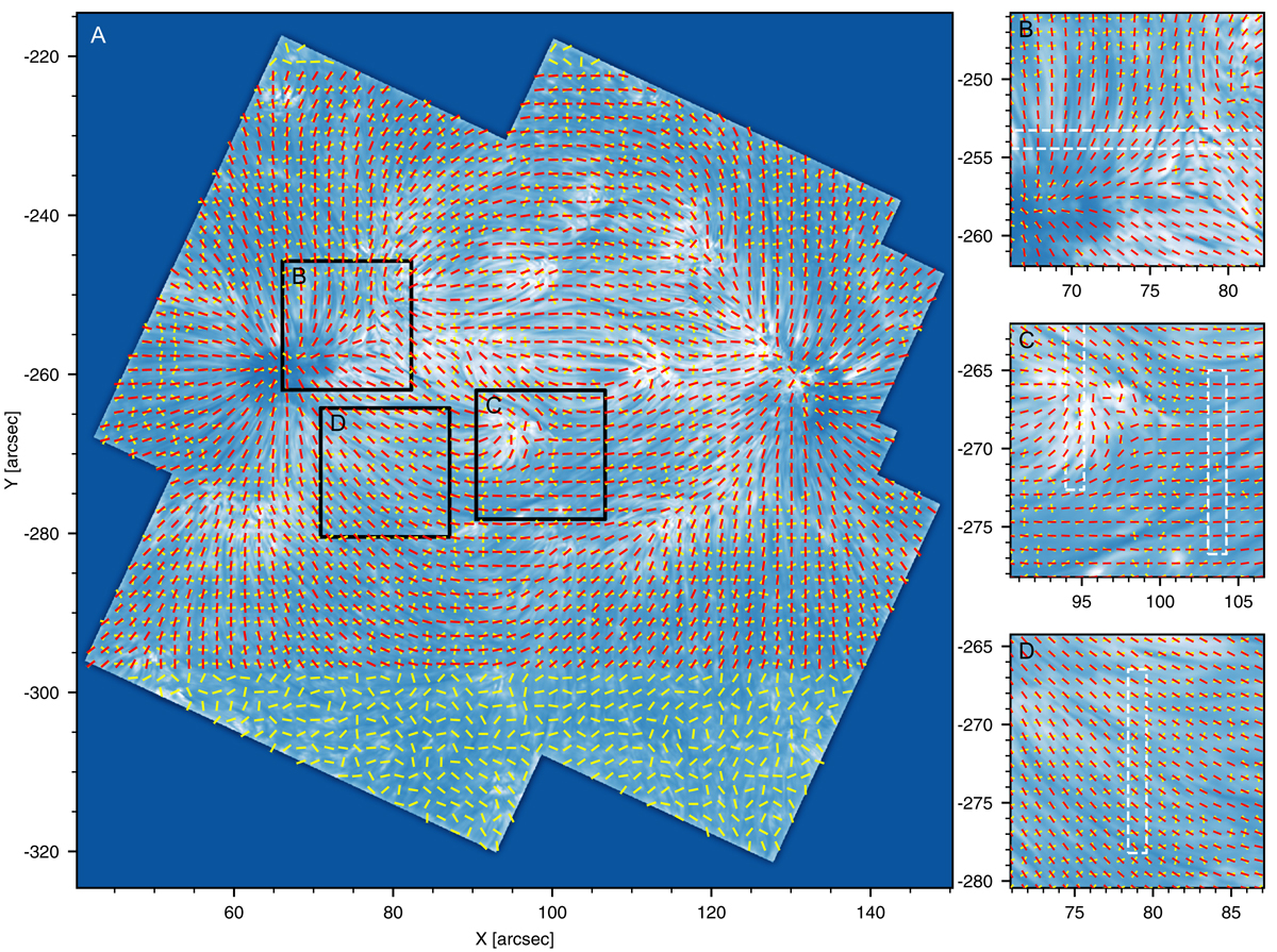
Comparison of fibril orientation and chromospheric field azimuth from the spatially-regularised WFA and the MHS model. The background Ca II 8542 Å image has been unsharp masked with a radius of 0![]() 5 to enhance fibrillar structures. The azimuth is indicated by yellow (WFA) and red (MHS) bars, where arrowheads have been omitted since we did not resolve the 180°-ambiguity for the WFA. The three white boxes in the left-hand panel a indicate the selections presented as panels b–d in the right-hand column and have been chosen such as to contain part of the ROIs in Fig. 1, where panel b covers ROI 1, panel c ROIs 2 and 3, and panel d ROI 4.
5 to enhance fibrillar structures. The azimuth is indicated by yellow (WFA) and red (MHS) bars, where arrowheads have been omitted since we did not resolve the 180°-ambiguity for the WFA. The three white boxes in the left-hand panel a indicate the selections presented as panels b–d in the right-hand column and have been chosen such as to contain part of the ROIs in Fig. 1, where panel b covers ROI 1, panel c ROIs 2 and 3, and panel d ROI 4.
Current usage metrics show cumulative count of Article Views (full-text article views including HTML views, PDF and ePub downloads, according to the available data) and Abstracts Views on Vision4Press platform.
Data correspond to usage on the plateform after 2015. The current usage metrics is available 48-96 hours after online publication and is updated daily on week days.
Initial download of the metrics may take a while.