Fig. 2
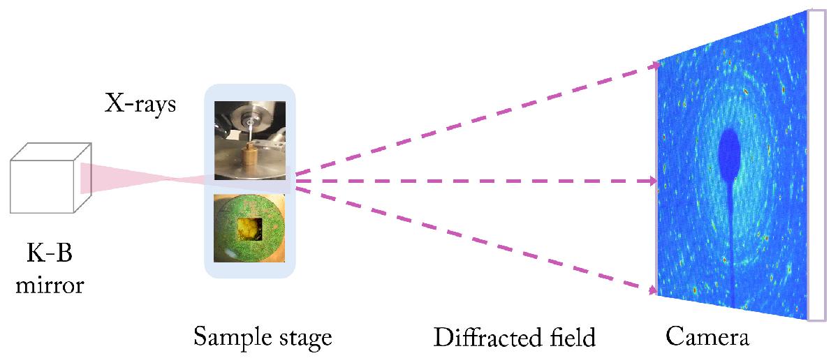
Schema of the powder diffraction setup at the ID16 beamline at the ESRF. X-rays are focused at the sample, consisting of polycrystalline MgSiO3 powder, placed in a capillary (φ = 780 μm) or powder deposited into a Si box (φ = 200 μm) holding a Si3N4 membrane. The transmission X-ray diffraction image is recorded behind the sample.
Current usage metrics show cumulative count of Article Views (full-text article views including HTML views, PDF and ePub downloads, according to the available data) and Abstracts Views on Vision4Press platform.
Data correspond to usage on the plateform after 2015. The current usage metrics is available 48-96 hours after online publication and is updated daily on week days.
Initial download of the metrics may take a while.