Fig. 3
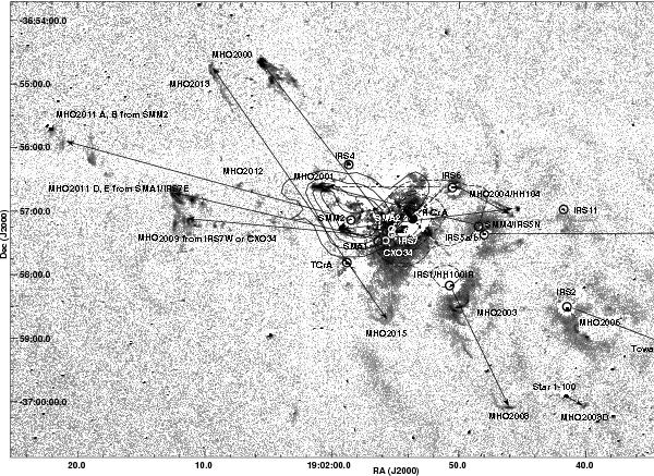
Continuum-subtracted H2 image of the Coronet subregion displayed using a logarithmic scaling. The circular symbols show all sources detected by the Spitzer IRAC images. The identifications are reproduced from Wilking et al. (1997) (IRS), Groppi et al. (2007) (SMA), and Nutter et al. (2005) (SMM). The arrows join the probable driving source with the corresponding MHO. When two arrows originate in the same source, it implies both outflow lobes are visible. The CO bipolar outflow mapped by Groppi et al. (2004) is shown by solid (blue lobe) and dotted (red lobe) contours. For the sake of clarity, MHO 2000, which is situated close to SMM2, is not marked in this figure.
Current usage metrics show cumulative count of Article Views (full-text article views including HTML views, PDF and ePub downloads, according to the available data) and Abstracts Views on Vision4Press platform.
Data correspond to usage on the plateform after 2015. The current usage metrics is available 48-96 hours after online publication and is updated daily on week days.
Initial download of the metrics may take a while.