Fig. 1
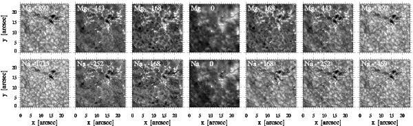
Profile-sampling image comparisons between the Mg line (upper row) and the Na line (lower row). These images are subfield cutouts from a dual-line SST/CRISP profile scan taken on August 23, 2010. They sample a small active region with pores in the upper part and a quiet area in the lower part of the subfield. Each image is byte-scaled independently and is labeled with its wavelength separation Δλ from line center in mÅ. Both sequences sample the line profile from the outer blue wing through the core to the outer red wing. The Δλ values in the first two columns were selected for scene similarity between the two lines. The next two columns were also selected for scene similarity but have equal Δλ values for the two lines. The final three columns mirror the Δλ values of the first three. All images show rich detail; we invite the reader to magnify them with a pdf viewer. This paper explains the imaging asymmetry between the two lines in the red-wing panels where the Mg line shows reversed granulation as in its blue wing, whereas the Na line instead shows normal granulation as in the last panel.
Current usage metrics show cumulative count of Article Views (full-text article views including HTML views, PDF and ePub downloads, according to the available data) and Abstracts Views on Vision4Press platform.
Data correspond to usage on the plateform after 2015. The current usage metrics is available 48-96 hours after online publication and is updated daily on week days.
Initial download of the metrics may take a while.