Fig. 4

Download original image
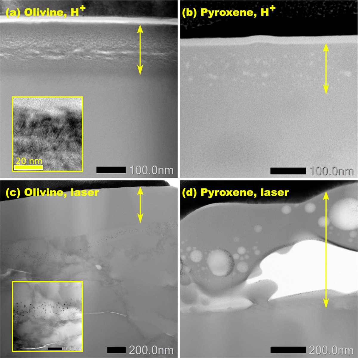
Bright-field scanning transmission electron microscopy images of the lamellae of all samples. Inset in (a) is the high-resolution transmission electron microscopy image of the altered layer in the H+-irradiated sample. Inset in (c) is the zoomed-in picture of the area containing nanophase Fe particles. Double-headed arrows mark the areas that are partially or fully amorphised.
Current usage metrics show cumulative count of Article Views (full-text article views including HTML views, PDF and ePub downloads, according to the available data) and Abstracts Views on Vision4Press platform.
Data correspond to usage on the plateform after 2015. The current usage metrics is available 48-96 hours after online publication and is updated daily on week days.
Initial download of the metrics may take a while.


