Figure 2 depicts the setup for the CONICA internal phase diversity measurements. The leftmost component carries the four pinholes, which are shifted against each other, with the values given above. In rotating the wheel holding the Zernike tool we are able to select a pinhole in the field of view. After a collimating lens and a pupil stop, a filter selects the wavelength range and finally the camera objective forms the object image on the detector. The chosen camera objective determines the f-ratio and the pixel scale.
To center the image of the pinholes on the detector, the whole pinhole
mount is shifted by turning the mask wheel. In principal PD needs the
input images to be on the same spot to ensure that the same
aberrations are sensed. The horizontal position of the pinholes can be
controlled by adjusting the rotation angle of the wheel. In vertical
direction there is no degree of freedom, but the four pinholes are
mounted circularly to compensate for the circular movement. By this
means a vertical precision of 50 mas (C50S) can be reached![]() . This is easily sufficient not
to see any influence due to field aberration effects. PD measurements
taken at different detector positions and calculations performed with
an optical design software showed that even at the corner of the field
of view (13 arcsec) the field aberration is negligible
(Paper I). Note that for some measurements in Paper I an earlier
version of the Zernike tool was used with a design not optimized for
the circular movement of the pinholes. The worst separation that
could occur with the former Zernike tool was about 1.3 arcsec. But
even with this tool no relevant impact on the precision of wavefront
sensing was detected.
. This is easily sufficient not
to see any influence due to field aberration effects. PD measurements
taken at different detector positions and calculations performed with
an optical design software showed that even at the corner of the field
of view (13 arcsec) the field aberration is negligible
(Paper I). Note that for some measurements in Paper I an earlier
version of the Zernike tool was used with a design not optimized for
the circular movement of the pinholes. The worst separation that
could occur with the former Zernike tool was about 1.3 arcsec. But
even with this tool no relevant impact on the precision of wavefront
sensing was detected.
Apart from the fact that the Zernike tool with its pinholes at the entrance focal plane provides the required focus shifts, it is convenient that the required focus shifts do not depend on the camera objective (pixel scale) anymore. But note: defocusing by moving an object in the entrance focal plane does not correspond exactly to a defocus due to a shifted detector plane. An investigation of this effect is done in Paper I and turns out to be negligible.
To summarise this section: the PD input data to derive the total
CONICA internal aberrations are obtained by object defocusing in the
CONICA entrance focal plane. The object defocusing is realized by four
10 ![]() m pinholes at different axial positions. Note that since the
entrance focal plane of CONICA is located inside the cold cryostat,
aberrations accrued from the CONICA entrance window are not included
in this wavefront estimation.
m pinholes at different axial positions. Note that since the
entrance focal plane of CONICA is located inside the cold cryostat,
aberrations accrued from the CONICA entrance window are not included
in this wavefront estimation.
Now, we describe how the PD input images are obtained which are used to sense the wavefront aberrations of the whole instrument, i.e., the adaptive optics NAOS together with its infrared camera CONICA. In this case we can take advantage of the AO system's capabilities to itself introduce an adequate focus shift and thus there is no need for the implementation of a special tool or a modifaction of the design.
In the entrance focal plane of NAOS, which coincides with the VLT
Nasmyth focal plane, a calibration point source can be slid in and
imaged by CONICA. This point source is realized by the output of a
fiber with a diameter of 10 ![]() m fixed on a movable stage. On the
same stage a second source much larger in diameter (400
m fixed on a movable stage. On the
same stage a second source much larger in diameter (400 ![]() m) is
mounted. It is only seen by the WFS and serves as a reference source
to close the loop. This extended source is needed for technical
reasons. In the case of no atmospheric turbulence the more extended
source provides a much better feed-back signal to the WFS than the
small one. By this means the AO control loop is adjusted for any
aberrations emerging in the common path. To obtain the focus shift
affecting the entire instrument, we introduce the desired amount of
defocusing in the WFS path by moving the mirrors of the field
selector. During this process the loop is kept closed. Instantly, the
arising focus shift is detected by the WFS. Correspondingly, the real
time computer commands the DM to compensate for the detected
defocus. Finally, the spots on the Shack-Hartmann WFS are centered
again, but the defocus of the DM takes effect in the imaging path.
For a pure defocus the DM will take a parabolic shape. The maximum
achievable defocus by this method is limited by the DM's stroke and
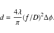
turns out to be about 20 mm. Refering to Table 1, this
is enough to introduce the needed diversity for an f/15 beam.
m) is
mounted. It is only seen by the WFS and serves as a reference source
to close the loop. This extended source is needed for technical
reasons. In the case of no atmospheric turbulence the more extended
source provides a much better feed-back signal to the WFS than the
small one. By this means the AO control loop is adjusted for any
aberrations emerging in the common path. To obtain the focus shift
affecting the entire instrument, we introduce the desired amount of
defocusing in the WFS path by moving the mirrors of the field
selector. During this process the loop is kept closed. Instantly, the
arising focus shift is detected by the WFS. Correspondingly, the real
time computer commands the DM to compensate for the detected
defocus. Finally, the spots on the Shack-Hartmann WFS are centered
again, but the defocus of the DM takes effect in the imaging path.
For a pure defocus the DM will take a parabolic shape. The maximum
achievable defocus by this method is limited by the DM's stroke and
turns out to be about 20 mm. Refering to Table 1, this
is enough to introduce the needed diversity for an f/15 beam.
The procedure is shown in Fig. 3 and provides us with the PD input data to estimate the NAOS-CONICA overall wavefront errors. In comparison with the procedure described in Sect. 3.1 we deal with the same object now, and we must not care about any deviations in the position of the image pairs. This simplifies data aquisition for the measurement and diminishes the number of possible error sources.
Copyright ESO 2003
![\begin{figure}
\par\includegraphics[width=8.8cm,clip]{fig/f2914_03.eps} \end{figure}](/articles/aa/full/2003/07/aa2914/img14.gif)