 |
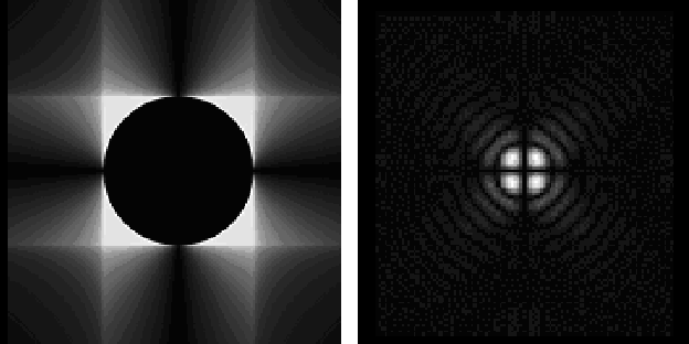
Figure 1: Theoretical intensity distribution in the pupil plane (left) and in the image plane (right) for a finite field of view. |
| Open with DEXTER | |
A&A 400, 385-392 (2003)
DOI: 10.1051/0004-6361:20021834
L. Abe1, 2 - A. Domiciano de Souza Jr.1, 2 - F. Vakili2 - J. Gay 1
1 - Observatoire de la Côte d'Azur, Département
Fresnel, UMR-CNRS 6528, 06460 Saint-Vallier de Thiey, France
2 - UMR-6525 Astrophysique, Université de Nice Sophia-Antipolis,
Parc Valrose, 06108 Nice Cedex 02, France
Received 28 February 2002 / Accepted 25 October 2002
Abstract
We report laboratory results from a monochromatic
prototype of the Phase Knife Coronagraph which validate our
previous theoretical and numerical simulations, prove the physical
principle of it and set realistic limitations to the nulling
properties of the coronagraph. The optical set-up, phase knife
manufacturing technique and different aspects of the instrumental
limitations are given. The first results attain easily a 3000
nulling effect obtained both on single and simulated double stars.
Optical and mechanical stabilities are discussed and future steps
to be carried out for a wide band version of this coronagraph are
outlined.
Key words: techniques: interferometric- methods: laboratory - stars: binaries: close
Unlike Lyot's coronagraph, interference nulling coronagraphs (Gay & Rabbia 1996; Roddier & Roddier 1997; Rouan et al. 2000) represent the only alternative to attain the resolution limit of present large telescopes like VLT (Monnet 2000), Keck telescopes (Wizinowich et al. 2000) and Gemini (Mountain et al. 1998). This have motivated our efforts to develop the Achromatic Phase-Knife Coronagraph (APKC hereafter, Abe et al. 2001, referred to as Paper I). In this paper we present the first laboratory results from PKC, the monochromatic version of the APKC, as a step along the way to construct a full-fledged high contrast imaging device to use on large ground-based monolithic telescopes equipped with extremely dense and high performance Adaptive Optics (A.O. hereafter, Mouillet 2001). In Paper I we described the conditions in which the intrinsic performances of PKC and its achromatic version APKC become similar. Therefore, in order to progress along our plan we have tested a monochromatic laboratory breadboard to validate its physical principle and to obtain realistic numbers on its operational characteristics. Section 2 summarizes the physical concept of the APKC. Section 3 describes phase knife manufacturing and commissioning, and the laboratory optical set-up used to assess its performances. Section 4 details the experimental results obtained on simple and binary simulated stars which settle the nulling performances of the APKC in its present configuration. Section 5 comments on the results and compares them to theory. In the final section we give a critical review of the APKC concept and show how future steps should bring us to a fully operational coronagraph for sky observations. The exact formalism for the residual energy inside the coronagraphic pupil is given in an appendix.
| |
Figure 1: Theoretical intensity distribution in the pupil plane (left) and in the image plane (right) for a finite field of view. |
| Open with DEXTER | |
It can be shown (see Appendix A) that in the case of an unobscured
telescope, with an infinite image plane phase mask and for a
monochromatic wave, the PKC completely rejects the on-axis source
energy outside of the conjugate pupil, offering in principle a
perfectly nulling coronagraph. Indeed the finite size of the
optical components (i.e. a finite field of view) in such a device
produces diffraction effects that re-inject energy inside the
conjugate pupil. Therefore, the final coronagraphic pattern
(Fig. 1, right) exhibits four bright
lobes resulting from diffraction residuals. Numerical simulations
show that for a
![]() field, and assuming perfect optics,
one can expect a reasonable 106 energy rejection for an
on-axis source.
field, and assuming perfect optics,
one can expect a reasonable 106 energy rejection for an
on-axis source.
In the real world however, imperfect optics, phase retardation properties, atmospheric turbulence and chromatic residues among other degrading effects limit the APKC nulling performances in ways that Sect. 3 will explicitly highlight.
In any high dynamic imaging process for exo-planet detection, the goal is to increase the local contrast between a central bright star (coronagraphic PSF) and its faint companion. Therefore, the energy rejection does not necessarily represent the best performance criterion. Hereafter we define the more appropriate, to our opinion, criteria to measure the coronagraphic efficiency used throughout this paper:
 |
(1) |
 |
(2) |
| = |  |
(3) | |
| = |  |
(4) |
This is the same problem as encountered with the Apodized Square Apertures (Nisenson 2001), the Dark Hole technique (Malbet 1995) and for those instruments that modify the final intensity distribution of a perfectly diffracted Airy pattern.
 |
Figure 2:
A phase knife sample seen through an interference microscope. The
step between the two fringe patterns from the phase knife zones
correspond to a geometrical phase shift which depends on the
aluminium deposit thickness (see text for further explanations).
The phase-shifts were measured using a 1 nm bandwidth
|
| Open with DEXTER | |
All tests were carried out with a full, unobscured pupil. The Lyot stop was chosen to be 70% of the full aperture in order to be less sensitive to optical tuning (stop centering) and residual low order aberrations such as defocus, which appears to be a major limitation to the coronagraph performance.
Although the two phase knives were built at the same time, they do
not produce the same phase-shift on the wave. This is interpreted
as being due to a non-uniform coating deposit inside the vacuum
tank. For the HeNe laser wavelength (632.8 nm), we measured the
phase-shift difference from the ideal ![]() value as
value as
![]() and
and
![]() which corresponds
respectively to a lack of thickness of
which corresponds
respectively to a lack of thickness of ![]() 5.5 nm and an excess
of thickness of
5.5 nm and an excess
of thickness of ![]() 0.5 nm. All measurements resulted from
correlations between the fringe patterns.
0.5 nm. All measurements resulted from
correlations between the fringe patterns.
The surface quality of the glass waveplates is locally good enough
(<![]() /50) for our application since we concentrate the light
on a very small area of the size of a few Airy discs. Indeed the
sharpness and rectilinearity of the step edge directly affect the
coronagraphic nulling performance, also limiting the practical
size of Airy patterns on the phase knives. The inspection of
interferograms showed that edge defects across a few tens of
millimeters remain below
/50) for our application since we concentrate the light
on a very small area of the size of a few Airy discs. Indeed the
sharpness and rectilinearity of the step edge directly affect the
coronagraphic nulling performance, also limiting the practical
size of Airy patterns on the phase knives. The inspection of
interferograms showed that edge defects across a few tens of
millimeters remain below
![]() at some locations,
and the phase transition edge itself extends over less than
at some locations,
and the phase transition edge itself extends over less than
![]() .
Indeed we used such optimum wave-plate zones as much
as possible.
.
Indeed we used such optimum wave-plate zones as much
as possible.
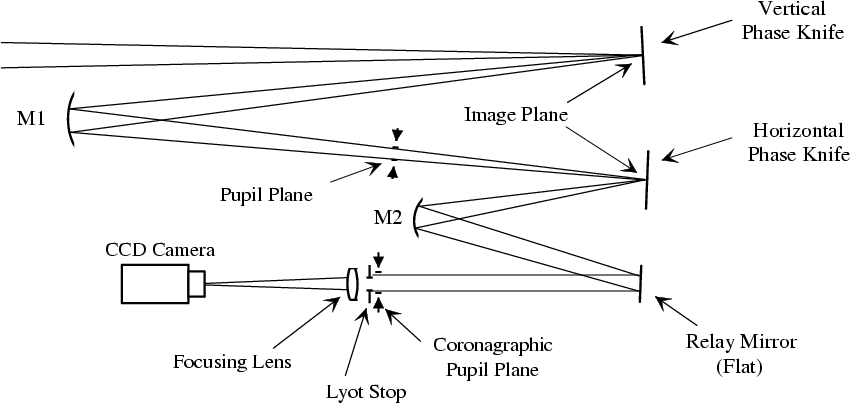 |
Figure 3: PKC optical layout. The two phase knives (horizontal and vertical) are equivalent to flat mirrors. M1 and M2 are spherical, forming the second focus and the final coronagraphic pupil image respectively. |
| Open with DEXTER | |
The pin-hole used for the source has a diameter of 10 microns.
With the collimating lens, the source is partially resolved by a
factor
![]() with respect to the Airy radius, roughly
corresponding to a 3 mas star observed on a 10 meters telescope in
the K band (
with respect to the Airy radius, roughly
corresponding to a 3 mas star observed on a 10 meters telescope in
the K band (
![]() ). Note that a partially resolved
object can dramatically affect the nulling performance of any
coronagraphic device in general and the APKC in particular.
). Note that a partially resolved
object can dramatically affect the nulling performance of any
coronagraphic device in general and the APKC in particular.
Figure 4 (bottom) shows the intensity distribution in the
final coronagraphic pupil plane obtained by numerical simulation
(right) and obtained under present laboratoy conditions (left). We
first note the remarkable reproduction of theory by experimental
results proving the physical validity of the PKC concept. From
this figure one could conclude that the measured rejection should
be extremely high but this is due to the CCD intensity dynamic
range limitation.
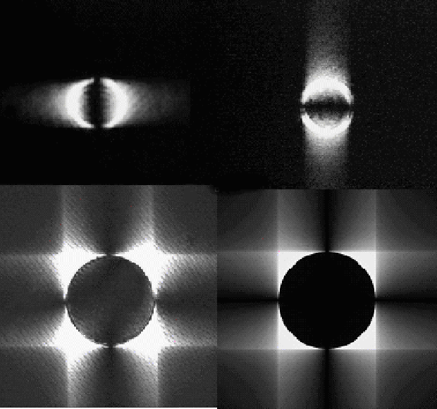 |
Figure 4: Comparison of actual laboratory and theoretical intensity distribution in successive pupil planes of the PKC coronagraph (not same scales): (top-left) pupil intensity after the vertical phase-knife alone, (top-right) pupil intensity after the horizontal phase-knife alone, (bottom-left) pupil intensity after both phase-knives being applied and (bottom-right) theoretical pupil intensity distribution. Note that for the actual pupil intensities the CCD exposure has been saturated to show the depth of the null inside the coronagraphic pupil. Fringes on the experimental data are due to multiple reflections on the CCD window. |
| Open with DEXTER | |
As already noted in Sect. 2.2, and since
we could not monitor the pupil image and the PSF simultaneously,
we decided to work on the final coronagraphic image rather than
measuring rejection rates in the pupil plane.
Figure 5 illustrates the nulling effect of the PKC
on a binary source. All three images have the same dynamic range
with an exposure time of 20 ms. The image on the left shows the
normal, i.e. without the PKC, image of a binary source. Placing
only one phase knife on the central source (middle) attenuates its
intensity already by a factor ![]() 10 as predicted by theory
(corresponding pupil intensity distributions are shown in
Fig. 4 (top)). In the right image, the combination of
both phase knives almost completely nulls the central bright
source. Due to the limited CCD dynamic range (8 bits) for a single
exposure the signal to noise ratio is not high enough to correctly
assess the coronagraphic PSF. Note also that the binary component
remains unaffected by the coronagraph at this location. According
to Paper I, for an off-axis companion falling exactly on the edge
of one of the two PKC knife edges, the effect would be identical
to the one of the bright source in the middle image of
Fig. 5.
10 as predicted by theory
(corresponding pupil intensity distributions are shown in
Fig. 4 (top)). In the right image, the combination of
both phase knives almost completely nulls the central bright
source. Due to the limited CCD dynamic range (8 bits) for a single
exposure the signal to noise ratio is not high enough to correctly
assess the coronagraphic PSF. Note also that the binary component
remains unaffected by the coronagraph at this location. According
to Paper I, for an off-axis companion falling exactly on the edge
of one of the two PKC knife edges, the effect would be identical
to the one of the bright source in the middle image of
Fig. 5.
| |
Figure 5: Application of the two-step PKC phase-knives to a laboratory binary source: left) the binary star star without coronagraph, middle) effect of the first horizontal phase-knife on the central bright star and right) the on-axis star being nulled after the second vertical phase-knife is applied. Images are shown with a non-linear intensity scale to enable direct comparison with the background noise. |
| Open with DEXTER | |
When removing the density filter, the coronagraphic PSF was still bright enough to match the CCD dynamic range for a short exposure of 20 ms. Consequently, it was not necessary to carry out time-consuming long exposures to attain the desired SNR. We could also monitor the effects of the tip-tilt jitter on the coronagraphic PSF while recording a series of short exposures. Indeed, the temperature inside the laboratory was relatively stable and produced only low order phase aberrations, using Zernike polynomials terminology, resulting in fast variations of both shape and intensity of the coronagraphic PSF.
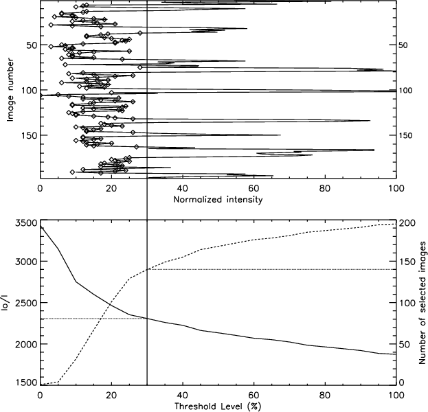 |
Figure 6:
(top) Coronagraphic PSF intensity maxima recorded during
a continuous series of 199 exposures (pixel intensity digitized
over 8 bits). (bottom) Image selection procedure applied to the
exposure set according to a flux threshold defined as a percentage
of the maximum intensity variation (full dynamic range of top).
The solid curve is the peak-to-peak intensity ratio (the
extinction, noted I0/I) between the off-axis reference
Airy pattern and the integrated coronagraphic PSFs selected by
applying the threshold. The dashed curve gives the number of
selected images after thresholding. In the given example, the 140
selected images are marked with diamonds on the top figure for a
threshold level of 30%, resulting in a long exposure extinction
of |
| Open with DEXTER | |
We applied various selection criteria to the image sequence
(Fig. 6, bottom) to determine how the overall extinction
is affected by "bad'' images (i.e. with low extinction). The
average extinction ratio for the long exposure for these 199
frames is 1870. Selecting half the images in the sequence results
in a ![]() 40% gain for the extinction. Results show that we can
increase the extinction up to a factor 2 in the best case.
However, the optimum selection criterion results from a good
compromise between rejection rate and the signal to noise ratio
for a possible off-axis object.
40% gain for the extinction. Results show that we can
increase the extinction up to a factor 2 in the best case.
However, the optimum selection criterion results from a good
compromise between rejection rate and the signal to noise ratio
for a possible off-axis object.
In the scope of determining the coronagraph's sensitivity to both phase-shift defects and tip-tilt jitter, we tried to reproduce the experimental conditions with a numerical simulation.
In a first step, we focused on pure optical and turbulence aberrations considering perfect phase knives: static optical aberrations derived from ray-tracing analysis, and numerical simulations show no significant effect on the final coronagraphic extinction. We conclude that the main degrading aberration is a systematic de-centering (constant shift) of the central source.
Then for turbulence aberrations we introduced tip-tilt jitter
derived from the off axis reference PSF measurements: we used a
Gaussian statistic with a 1% rms amplitude in ![]() units
(see Sect. 4.3) and a mean position exactly
centered the coronagraph's axis. The phase knives were first
considered perfectly
units
(see Sect. 4.3) and a mean position exactly
centered the coronagraph's axis. The phase knives were first
considered perfectly ![]() -shifting. The same image selection
procedure as for the experimental data was used, as shown in
Fig. 7 (top). This graph indicates that
only
-shifting. The same image selection
procedure as for the experimental data was used, as shown in
Fig. 7 (top). This graph indicates that
only ![]() 10% of the images have a very good extinction,
greater than
10% of the images have a very good extinction,
greater than
![]() ,
with a maximum at
,
with a maximum at ![]() 106 as
predicted by the perfect static model.
106 as
predicted by the perfect static model.
Now, when introducing the measured phase-shift defects on both
knives, and using the same image selection
(Fig. 7, bottom), the expected maximal
extinction is ![]() 3370, with a long exposure extinction of
3370, with a long exposure extinction of
![]() 3070. This significant loss of performance is rather close
to our experimental results. We also notice that the proportion of
images with a good extinction is much higher and always very close
to the maximum extinction which means that the extinction variance
(in percentage) is much lower than in the perfect case.
3070. This significant loss of performance is rather close
to our experimental results. We also notice that the proportion of
images with a good extinction is much higher and always very close
to the maximum extinction which means that the extinction variance
(in percentage) is much lower than in the perfect case.
We conclude that the dominant degradation comes from imperfect phase-shift on the phase knives rather than the tip-tilt jitter.
 |
Figure 7:
(top) Image selection process of 200 numerically
computed images where the experimental conditions were introduced
(tip-tilt jitter and phase knives defects). (bottom) Same as top
with tip-tilt jitter only. The Airy spot is perfectly centered on
the coronagraph's axis with a gaussian tip-tilt jitter of 1% rms
( |
| Open with DEXTER | |
Results of Fig. 7 (top) are better than
the experimental ones (Fig. 6), but the assumption we
made of a perfectly centered spot on the coronagraph axis is a too
restrictive hypothesis, since we could not verify it during the
experiments. By introducing a centering error (in this case for
example 8% of ![]() in the vertical direction), we can
almost reproduce the same statistics
(Fig. 8) and intensity distribution
(Fig. 9). Such an error on the
centering appears to be rather large compared to the manual tuning
precision of each individual optical element (the two phase
knives). Nevertheless, there are other tuning error sources (e.g.
turbulence instabilities, visual estimations of the residual flux,
separate manual tuning for each phase knife) that we could not
monitor in real-time. Therefore we point out that for an optimized
PKC one requires an extremely accurate and robust tuning
procedure.
in the vertical direction), we can
almost reproduce the same statistics
(Fig. 8) and intensity distribution
(Fig. 9). Such an error on the
centering appears to be rather large compared to the manual tuning
precision of each individual optical element (the two phase
knives). Nevertheless, there are other tuning error sources (e.g.
turbulence instabilities, visual estimations of the residual flux,
separate manual tuning for each phase knife) that we could not
monitor in real-time. Therefore we point out that for an optimized
PKC one requires an extremely accurate and robust tuning
procedure.
 |
Figure 8: Same curve as Fig. 7 but where a centering error of 8% of the Airy spot on the coronagraph axis has been introduced. |
| Open with DEXTER | |
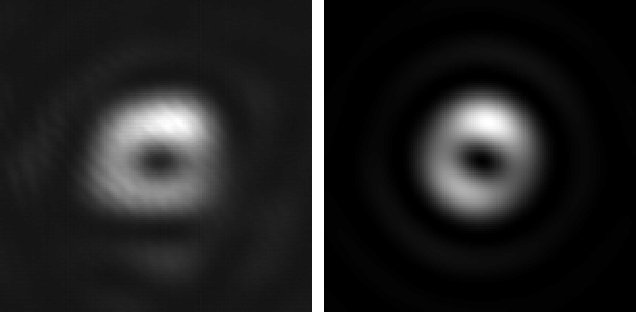 |
Figure 9:
Long exposure images of (left) the experimental data and
(right) simulated data, corresponding to the graph of
Fig. 8. Notice that the bright bump
location on top of the ring on both images almost exactly match.
It mainly originates from the important phase-shift defect
( |
| Open with DEXTER | |
This study also shows that the optimal phase knife manufacturing
quality depends on the observing strategy and AO/detector
capability: different tip-tilt jitter and phase-shift defect
combinations can result in an identical integrated extinction
(i.e. summing all exposures). For example, in our case, a 1.5%
rms tip-tilt jitter (![]() units) associated with perfectly
phase-shifting knives roughly gives the same integrated extinction
(
units) associated with perfectly
phase-shifting knives roughly gives the same integrated extinction
(
![]() )
as a 0.5% jitter and a 1.0% phase knife defect. On
the contrary, if an image selection strategy is chosen using a
very fast and sensitive detector (e.g. a photon-counting camera),
then top quality manufacturing will be mandatory to allow the
optimal performance of the coronagraph. Note that in this case if
the flux is very low, one needs to perform quite long exposures,
losing the possibility to access a statistical analysis of the
data in order to select the best images. Therefore the importance
of sensitive and fast read-out detectors for coronagraphic
instruments, such as fast photon counting cameras or new Low Light
Level CCDs (Mackay 2001; Jerram 2001) becomes more clear.
)
as a 0.5% jitter and a 1.0% phase knife defect. On
the contrary, if an image selection strategy is chosen using a
very fast and sensitive detector (e.g. a photon-counting camera),
then top quality manufacturing will be mandatory to allow the
optimal performance of the coronagraph. Note that in this case if
the flux is very low, one needs to perform quite long exposures,
losing the possibility to access a statistical analysis of the
data in order to select the best images. Therefore the importance
of sensitive and fast read-out detectors for coronagraphic
instruments, such as fast photon counting cameras or new Low Light
Level CCDs (Mackay 2001; Jerram 2001) becomes more clear.
These considerations should be taken into account for a future
APKC in order to significantly improve its performances relative
to the present operating prototype and to approach the theoretical
nulling effect of 106, specially for a wide-band version. The
phase knife manufacturing for the APKC concept as described in
Paper I differs from this achromatic version by the prismatic
shape to be given to the knives. The end-to-end rectilinearity of
the phase transition should be kept less than a few percent of
![]() /D as well as local edge defects. A more complete study
should be carried out in order to estimate the sensitivity to
these types of defects.
/D as well as local edge defects. A more complete study
should be carried out in order to estimate the sensitivity to
these types of defects.
The tip-tilt should be monitored independently of the main AO loop
using the rejected light outside the coronagraphic pupil. It
should be noted that fine tip-tilt compensation in APKC can be
achieved by equipping the two phase knives (see Fig. 2 of Paper I)
independently with a single fast linear motion actuator. For a 1
mm diameter Airy spot, such an actuator should have a typical
dynamic range of a few tens of microns with a linear resolution of
one micron corresponding to a tip-tilt fine compensation better
than
![]() .
On the other hand the combination of two
successive reflection phase-knives in APKC and the exact chromatic
phase-retardation compensation by means of diffraction gratings
can be applied for IR observations. This characteristic combined
with the dispersing/de-dispersing technique to obtain an
achromatic PKC represent a straightforward way to transform the
APKC to a spectrometric coronagraph. This is particularly adapted
to spectrally characterize an exoplanet once it has been detected
in the wide band configuration of APKC. At the present time we
have begun the commissioning of a hybrid version of the APKC and
the 4QC (Rouan et al. 2000). The coronagraph will be tested in sky
observation runs using a photon-counting camera and image
selection as described above. The exact coronagraph configuration
and the results will be discussed in a future paper.
.
On the other hand the combination of two
successive reflection phase-knives in APKC and the exact chromatic
phase-retardation compensation by means of diffraction gratings
can be applied for IR observations. This characteristic combined
with the dispersing/de-dispersing technique to obtain an
achromatic PKC represent a straightforward way to transform the
APKC to a spectrometric coronagraph. This is particularly adapted
to spectrally characterize an exoplanet once it has been detected
in the wide band configuration of APKC. At the present time we
have begun the commissioning of a hybrid version of the APKC and
the 4QC (Rouan et al. 2000). The coronagraph will be tested in sky
observation runs using a photon-counting camera and image
selection as described above. The exact coronagraph configuration
and the results will be discussed in a future paper.
We conclude that although a space-borne telescope appears as the
natural site to implement an APKC, a ground-based instrument
allows direct imaging of
![]() with realistic
turbulence and residual tip-tilt errors of
with realistic
turbulence and residual tip-tilt errors of ![]() 10% (Paper I,
Rouan et al. 2000). Benefiting from our laboratory results, the APKC
concept clearly constitutes a potential candidate for future
ground-based adaptive optics plus coronagraphic instruments for
exoplanet hunting similar to the VLT-Planet Finder foreseen in the
near future on an 8 m telescope.
10% (Paper I,
Rouan et al. 2000). Benefiting from our laboratory results, the APKC
concept clearly constitutes a potential candidate for future
ground-based adaptive optics plus coronagraphic instruments for
exoplanet hunting similar to the VLT-Planet Finder foreseen in the
near future on an 8 m telescope.
In the pupil plane, we express the spatial frequency coordinates
as:
As already mentioned in Paper I, the amplitude distribution is the
result of the convolution between the entrance pupil function and
the Fourier transform of the PKC function
![]() in the infinite field case
which can be written as,
in the infinite field case
which can be written as,
 |
(A.1) |
We express the integration process in the sense of the principal
value by excluding from the integration domain the symmetric areas
surrounding the discontinuities as shown in
Fig. A.1. The radius
![]() and the
angle
and the
angle ![]() respectively prevent divergence at the center point
and along the
respectively prevent divergence at the center point
and along the ![]() and
and ![]() axis. These parameters will tend to
zero once the result of the convolution product is established.
axis. These parameters will tend to
zero once the result of the convolution product is established.
Using Eq. (A.2), the convolution product
![]() is written
is written
| A(u,v) | = |  |
|
| = | ![$\displaystyle -\frac{2} {{\pi ^2 }}\int\limits_{\vartheta} {\frac{1}
{{\sin(2\v...
...a)}}} \left[ \ln (C(\vartheta,u,v) - \ln
(\varepsilon )\right]{\rm d}\vartheta.$](/articles/aa/full/2003/10/aa2428/img61.gif) |
| A(u,v) = | - | ![$\displaystyle \frac{2} {{\pi ^2 }}\int\limits_{\vartheta = \alpha
}^{\frac{\pi}...
...theta)}}}
\left\{
\left[ \ln(C(\vartheta,u,v) - \ln(\varepsilon) \right]\right.$](/articles/aa/full/2003/10/aa2428/img65.gif) |
|
| - | |||
| + | |||
| - |
 |
(A.4) |
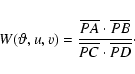
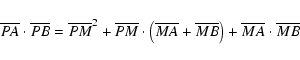
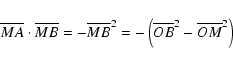
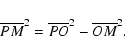
By a point P of the plan which contains a circle of center Oand radius R, we draw a line intersecting the circle at points
A and B. The perpendicular to AB passing through Ointersects the segment AB in its middle point M (Fig. B.1).
The product
![]() can be decomposed
as,
can be decomposed
as,

Acknowledgements
L. Abe is grateful to R. Krawczyk and Alcatel-Space Industries/Cannes for supporting his PhD fellowship. A.Domiciano de Souza Jr. acknowledges CAPES - Brazil (contract BEX 1661/98-1) for financial support. APKC development is supported by the ASHRA-CNRS program in France. The authors would like to thank the following people for their assistance in the APKC laboratory prototype realization: P. Assus, Y. Bresson, F. Desenfant, A. Glintzlin, J.-L. Schneider, A. Spang and N. Thureau. This paper benefitted from critical discussions with C. Aime, L. Arnold, D. Bonneau, A. Labeyrie, D. Mourard, Y. Rabbia, R. Soummer, E. Thomas and D. Vernet.

![\begin{figure}
\par\includegraphics[width=7cm, clip]{ABE2428f12.eps}\end{figure}](/articles/aa/full/2003/10/aa2428/img53.gif)


