 |
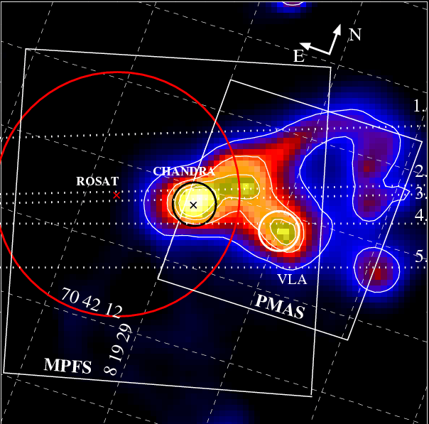
Figure 1:
CFHT archival H |
| Open with DEXTER | |
In the text
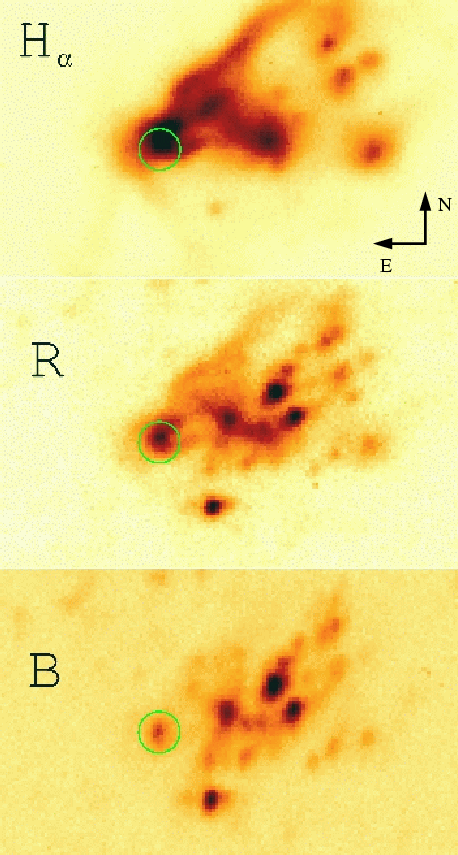 |
Figure 2:
CFHT H |
| Open with DEXTER | |
In the text
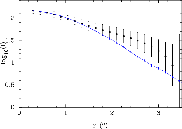 |
Figure 3:
Surface brightness profile of the He II emission around the ULX (solid
circles) together with the MPFS PSF (solid line). The error bars include
both the photon noise and the error in the surface brightness
determination. The He II region around the ULX is clearly extended to
about 3
|
| Open with DEXTER | |
In the text
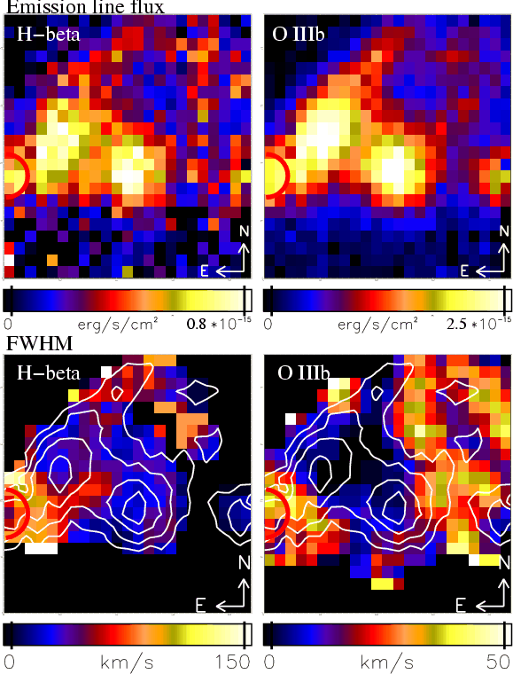 |
Figure 4:
PMAS emission line flux maps ( upper panels), and emission line FWHM maps
( lower panels), corrected for instrumental resolution, for H |
| Open with DEXTER | |
In the text
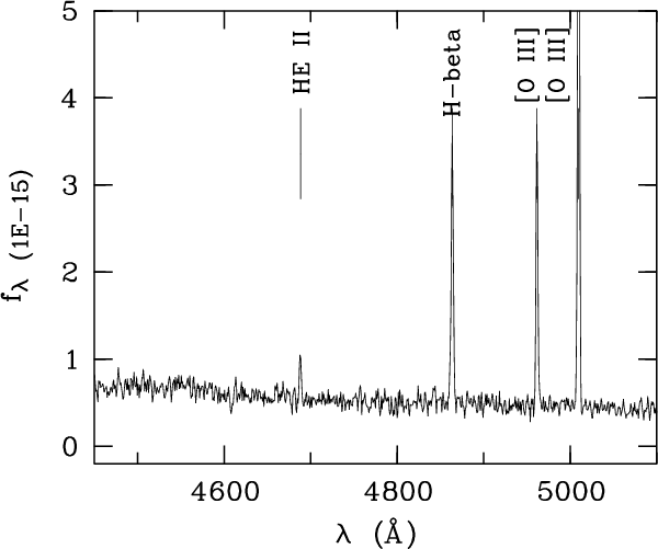 |
Figure 5:
The co-added PMAS spectrum at the X-ray position (see Fig. 4) clearly shows an He II
|
| Open with DEXTER | |
In the text
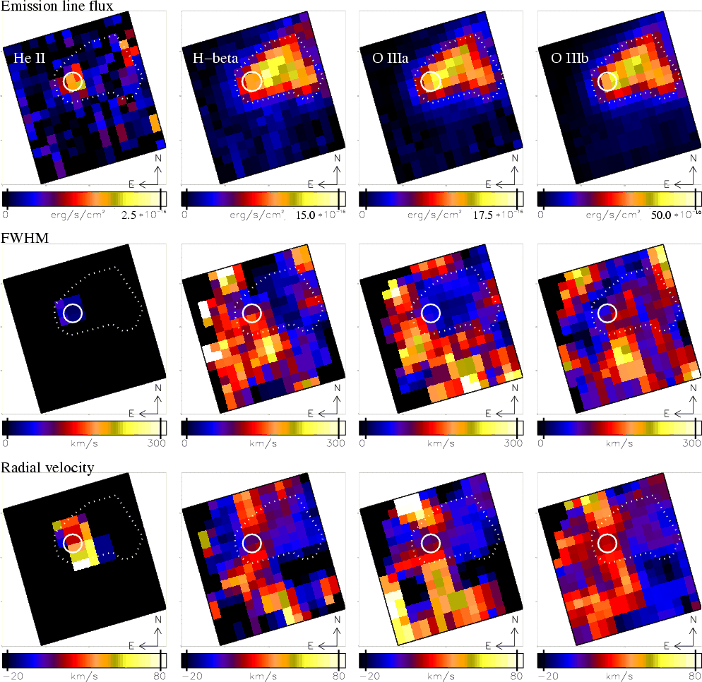 |
Figure 6:
MPFS emission line flux, FWHM (corrected for instrumental resolution) and radial velocity maps for He II, H |
| Open with DEXTER | |
In the text
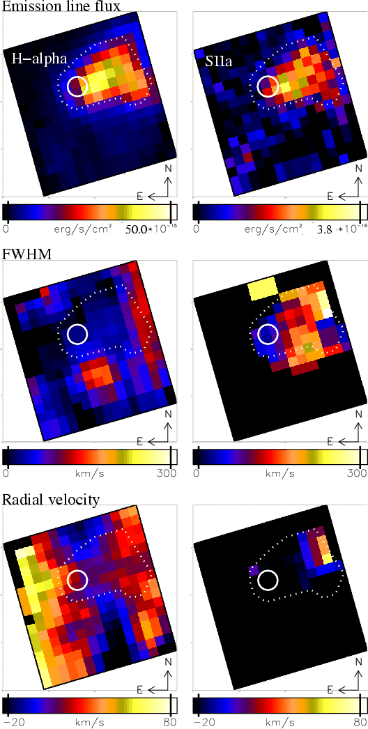 |
Figure 7:
Same maps as shown in Fig. 6 for H |
| Open with DEXTER | |
In the text
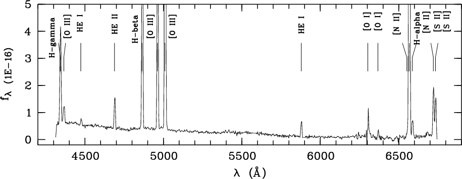 |
Figure 8:
The average LSS spectrum of the slit positions 2 and 3 (see Fig. 1) at the X-ray source shows, in addition to the lines found
in the MPFS spectra, faint [O III]
|
| Open with DEXTER | |
In the text
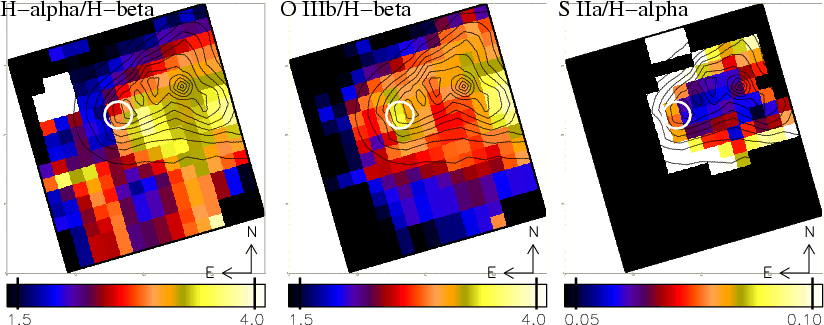 |
Figure 9:
MPFS line flux ratio maps of Ho II X-1: H |
| Open with DEXTER | |
In the text
| |
Figure 10:
Monochromatic MPFS images in He II
|
| Open with DEXTER | |
In the text
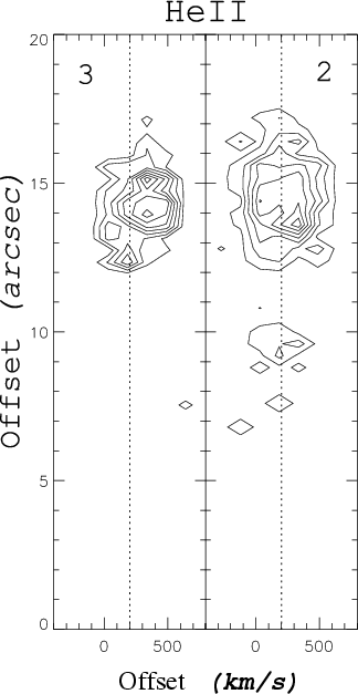 |
Figure 11:
Isophotes of the 2d-LSS spectra at slit positions N2 and N3 in the He II
|
| Open with DEXTER | |
In the text
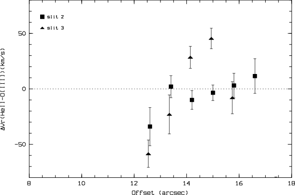 |
Figure 12:
Radial velocity difference derived from the HeII
|
| Open with DEXTER | |
In the text
![\begin{figure}
\par\includegraphics[angle=90,width=8.5cm,clip]{0827fg13.ps}\hspace*{3mm}
\includegraphics[width=6cm,height=6.3cm,clip]{0827fg14.ps}
\end{figure}](/articles/aa/full/2005/09/aa0827/img143.gif) |
Figure 13:
Left: radial profile of Holmberg II X-1 (diamonds with error bars) compared to the simulated ACIS-S PSF at 1 keV. A pixel corresponds to 0.5
|
| Open with DEXTER | |
In the text
![\begin{figure}
\par\includegraphics[width=7.9cm,clip]{0827fg15.eps}
\end{figure}](/articles/aa/full/2005/09/aa0827/img201.gif) |
Figure 14:
The X-ray pulse-height spectrum of XMM EPIC-PN
is shown with folded models for fit (B).
The lower panel shows the data/model ratios. Pulse height spectra
are re-binned for display and 1 |
| Open with DEXTER | |
In the text
![\begin{figure}
\par\includegraphics[width=8cm,clip]{0827fg16.eps}
\end{figure}](/articles/aa/full/2005/09/aa0827/img202.gif) |
Figure 15: Unfolded XMM EPIC-PN spectrum showing the best fit model (B). The thermal black body component contributes about 18% to the total 0.3-8.0 keV flux, and is clearly soft (<2 keV). |
| Open with DEXTER | |
In the text